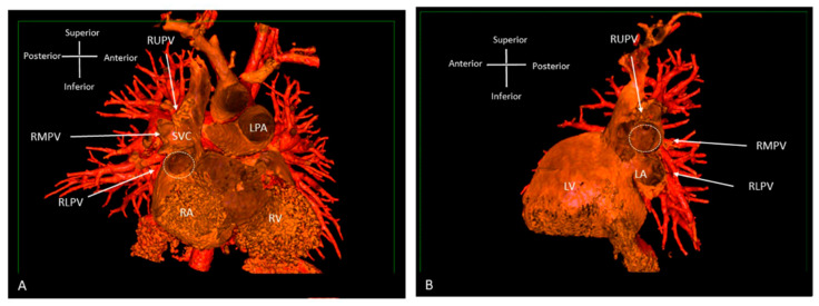
Figure 8.
Three-dimensional rendered images from CT, demonstrating the appearance of Superior Sinus Venosus ASD. (A) Demonstrates a superior sinus venosus ASD as outlined by the dashed line—seen from the right atrial aspect. RUPV and RMPV drain anomalously to the SVC. RLPV can be seen draining to LA. (B) Depicts a superior sinus venosus ASD from the left atrial aspect, allows imaging of the anomalous drainage of the RUPV and RMPV to the SVC. RLPV illustrated draining into the LA. LA: Left atrium; LPA: Left pulmonary artery; LV: Left ventricle; RA: Right atrium; RV: Right ventricle; SVC: Superior vena cava; RLPV: Right lower pulmonary vein; RMPV: Right middle pulmonary vein; RUPV: Right upper pulmonary vein.

