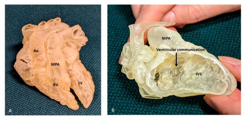
Figure 10.
Three-dimensional printed model from a case of DORV. (A) View of the anterior surface of the heart, demonstrating the relation of the great arteries, both committed to RV. (B) Right ventricular view of interventricular communication, looking at the IVS “en face”. The rims of the defect can be appreciated, but there is paucity of any additional intracardiac details, especially with regard to TV. Ao: Aorta; LV: Left ventricle; MPA: Main pulmonary artery; RV: Right ventricle.

