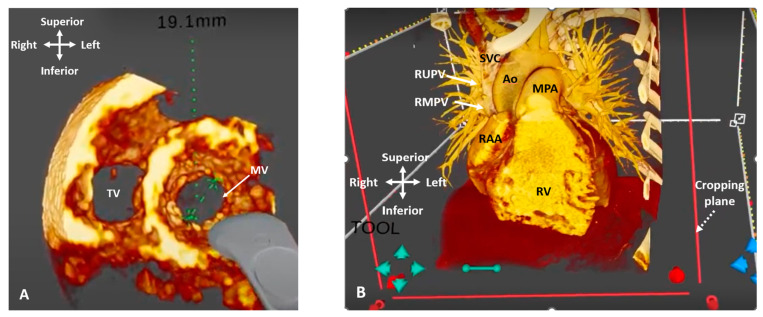
Figure 13.
Three-dimensional rendered imaging in virtual reality. (A) 3D TOE image viewed from ventricular apex, demonstrating view of the mitral and tricuspid valves. A measurement of the mitral valve annulus has been performed using the in-built tool. (B) CT demonstrating a case of Superior Sinus Venosus ASD. The cropping plane (dashed arrow) is cropping into the volume from the anterior plane and demonstrating the entry points of the RUPV and RMPV into the SVC (arrows). Ao: Aorta; MPA: Main pulmonary artery; MV: Mitral valve; RAA: Right atrial appendage; RMPV: Right middle pulmonary vein; RUPV: Right upper pulmonary vein; RV: Right ventricle; SVC: Superior vena cava; TV: Tricuspid valve.

