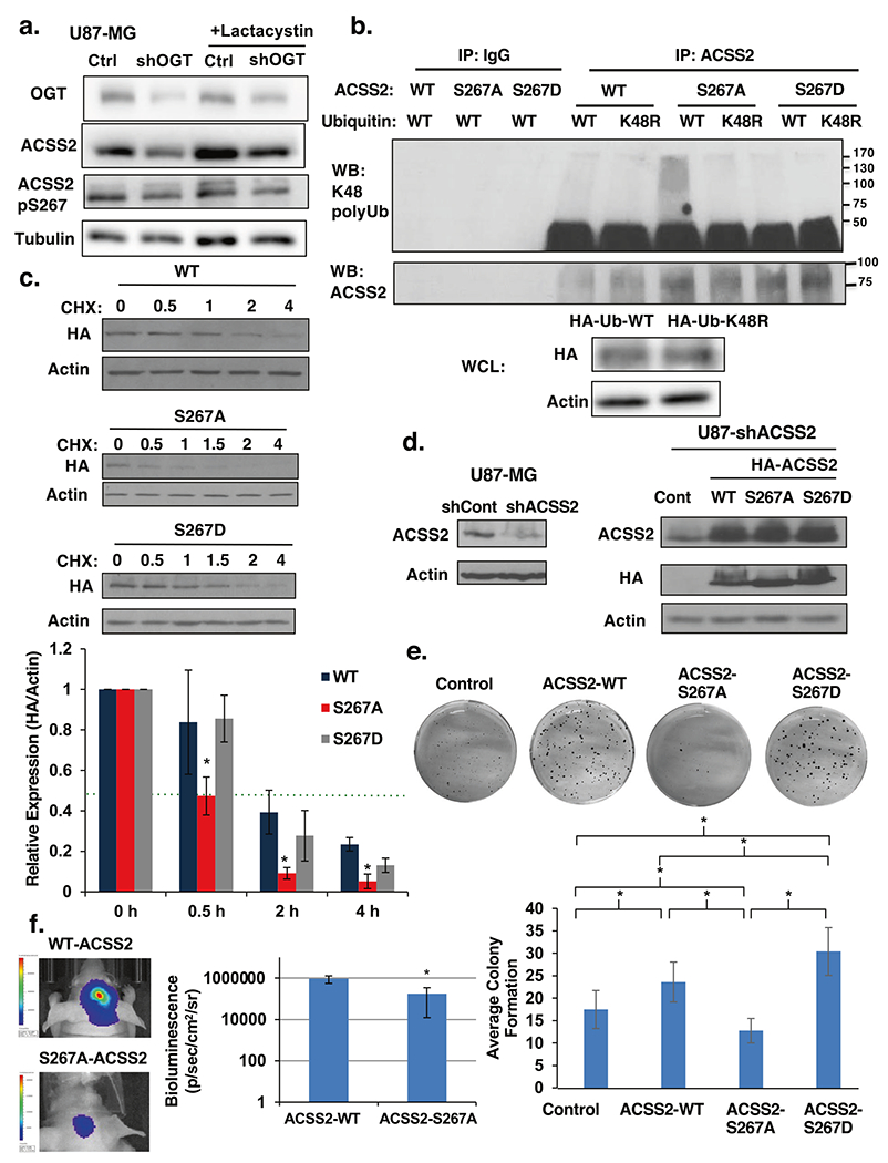
Fig. 5. Phosphorylation of Ser267 enhances stability of ACSS2 and is required for GBM growth.

a Cell lysate from U87-MG cells stably expressing control or OGT shRNA and treated with the proteasomal inhibitor 10 μM lactacystin for 6 h were collected for immunoblot analysis with the indicated antibodies. b Immunoprecipitation was performed with the indicated antibodies from U87-MG cell lysates stably expressing wild-type (WT)-, S267A-, or S267D- HA-ACSS2 and transfected with Ub-WT or Ub-K48. c Cell lysate from U87-MG cells stably expressing wild-type (WT)-, S267A-, or S267D-HA-ACSS2 in U87-MG glioblastoma cells treated with 10 μg/μl cycloheximide for indicated time (hours) were collected for immunoblot analysis with the indicated antibodies (top). Densitometry quantification of three independent time-course experiments presented as relative to expression (HA/actin) to 0 h (bottom). Green dotted line represents 0.5 relative expression. Student’s t-test reported as mean ± SD. *p-value < 0.05. d Cell lysates from U87-MG cells stably expressing ACSS2 shRNA against endogenous 3′ UTR of ACSS2 (left) and stably overexpressing wild-type ACSS2, ACSS2-S267A and ACSS2-S267D mutant (right) were collected for immunoblot analysis with the indicated antibodies. e Representative image of cells in d seeded into an anchorage-independent growth assay and imaged at day 14 (top). Data are quantified and presented as average from at least three independent experiments (bottom). Student’s t-test reported as mean ± SD. *p-value < 0.05. f Representative images of tumor growth detected via bioluminescence at Day 16 following injection of U87-MG-luciferase cells WT-ACSS2 or S267A-ACSS2 (left). Quantification of tumor size (WT n = 4, S267A n = 4) (right). Student’s t-test reported as mean ± SD. *p-value < 0.05.
