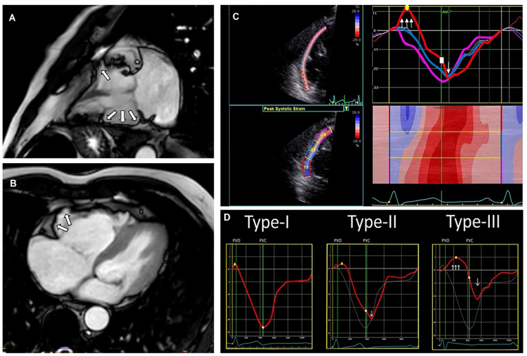
Figure 4.
Cardiac magnetic resonance (CMR) images from a patient with arrhythmogenic cardiomyopathy (ACM), including a typical echocardiographic deformation imaging pattern. CMR images (A,B) with right ventricular (RV) aneurysm (white arrows). Echocardiographic deformation imaging (C) with RV focused and peak systolic strain measurements and deformation pattern. Distinct types of RV deformation patterns (D) as determined by Mast et al. [47]; type-I: normal pattern; type-II: “delated onset of shortening and decreased systolic peak strain”; type-III: “systolic stretching and passive recoil or shortening during early diastole”. The patients’ strain pattern shows a type-III strain pattern, fitting structural ACM.

