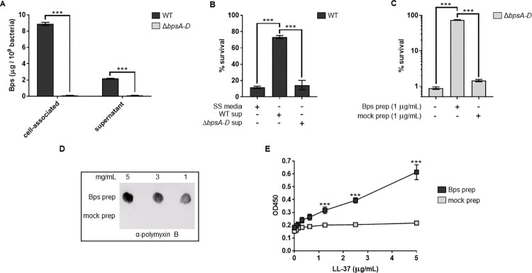
Fig 4. B. pertussis releases Bps during laboratory growth, and cell-free Bps provides protection from and binds to PmB and LL-37.
(a) Quantitation of cell-associated and released (supernatant) Bps from the WT and ΔbpsA-D strains by ELISA. Each data point represents the mean and s.e.m. of eight wells from one experiment and is representative of two independent experiments. Statistical differences were assessed by two-way ANOVA. ***, p<0.0005. (b, c) Survival of WT and ΔbpsA-D strains in the presence of PmB (b) or LL-37 (c). Supernatants from WT and the ΔbpsA-D cultures (b) or purified Bps and mock preparation (c) were added as indicated. Each data point represents the mean and s.e.m. of triplicates from one experiment and is representative of two independent experiments. Statistical differences were assessed by one-way ANOVA. ***, p<0.0005. (d, e) Bps binds PmB and LL-37. (d) Bps or mock preparations were spotted on nitrocellulose membranes. To detect PmB binding, the membranes were incubated with PmB, washed and probed with α-polymyxin B antibody conjugated to HRP. (e) Binding of Bps or mock preparations to LL-37 was quantified by ELISA using WGA conjugated to HRP. Asterisks indicate significance compared to mock prep. Each data point represents the mean and s.e.m. from one experiment and is representative of two independent experiments. Statistical differences between Bps and mock preps were assessed by unpaired two-tailed Student’s t test. ***, p<0.0005.

