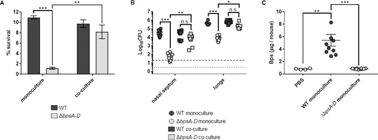
Fig 5. The presence of the WT strain increases ΔbpsA-D resistance to LL-37 in vitro and enhances its survival in the mouse respiratory tract and Bps is released in mouse lungs during infection.
(a) Survival of WT and ΔbpsA-D strains either in monoculture or in a 1:1 co-culture of both strains against LL-37. Each data point represents the mean and s.e.m. of triplicates from one of five independent experiments. Statistical differences were assessed by two-way ANOVA. **, p<0.005; ***, p<0.0005. n.s. not significant. (b) Bacterial CFUs recovered from the nasal septum and lungs of C57BL/6J mice four days after aerosol infection with WT or ΔbpsA-D in monoculture or in a 1:1 co-culture. Bars indicate the mean and s.e.m. of ten mice. Data from two independent experiments with groups of five mice each are shown. Statistical differences were assessed by two-way ANOVA. *, p<0.05; **, p<0.005; ***, p<0.0005; n.s., not significant. Dotted line represents the lower limit of detection for nasal septum, and dashed line represents the lower limit of detection for lungs. (c) Amounts of Bps in supernatants of lung lysates obtained from mice either instilled with PBS (blank circles) or infected with the indicated strains was quantified by ELISA using WGA conjugated to HRP. For infected mice, bars indicate the mean and s.e.m. of two independent experiments consisting of groups of five mice each (from Fig 5B). For PBS-instilled mice, bars indicate the mean and s.e.m. of one experiment consisting of four mice. Statistical differences were assessed by one-way ANOVA. **, p<0.005; ***, p<0.0005.

