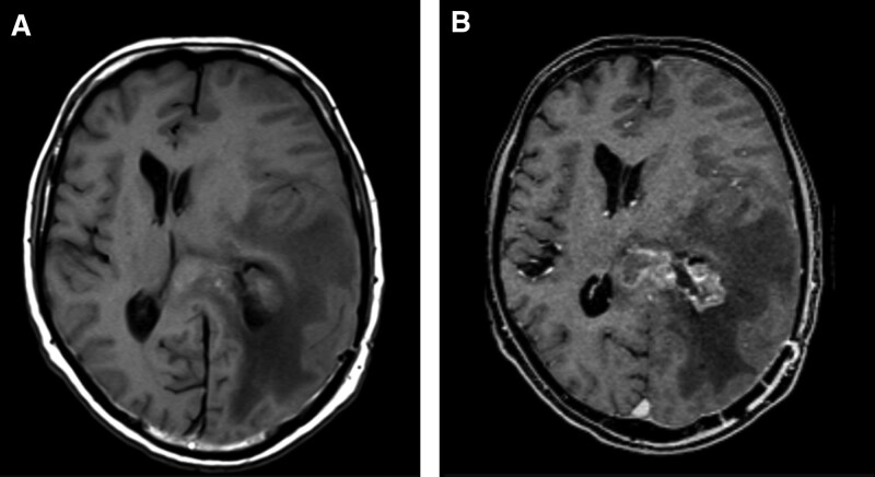
Figure 1.
High grade brain glioma pre (a) and post administration of contrast (b), in a 32-year-old male patient. A poorly defined Heterogeneous left parietal mass in T1 without contrast, associated with marked vasogenic edema and midline shift to the contralateral side. After administration of contrast (Clariscan, 0.10 mmol/kg), it shows heterogeneous enhancement and allows to distinctly define the border of the left periventricular parietal mass with ependymal extension with marked vasogenic edema and midline shift. Image quality was reported as good by site investigator.

