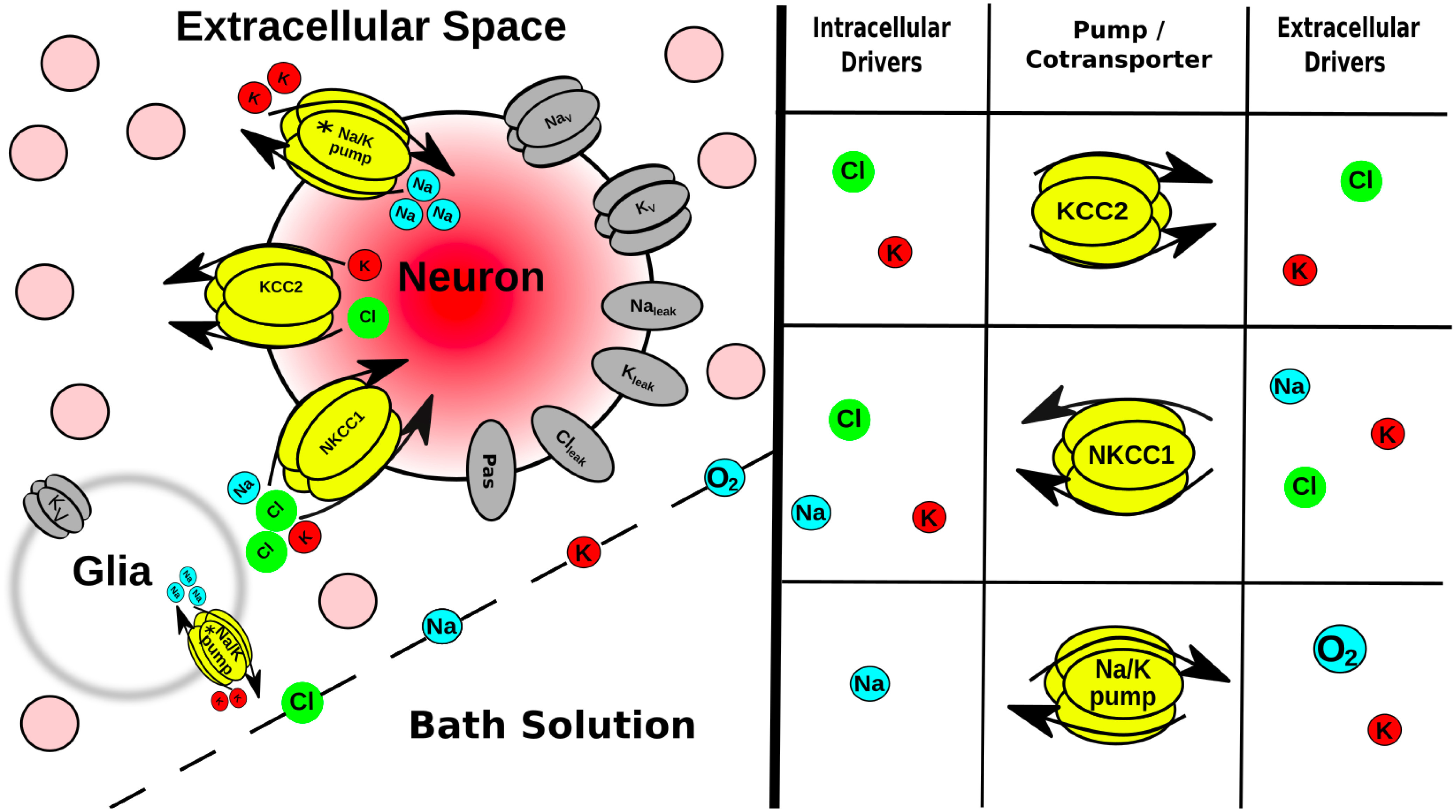
Figure 1.

Multiscale model expanded. Tissue scale: a few of the 36 ⋅ 103 neurons (pink circles) embedded in the ECS of a brain slice submerged in a bath solution where ion and O2 concentrations were held constant. Glia are not explicitly modeled, but instead were represented as a field of sinks in every ECS voxel. Cell scale: each neuron had ion channels, 2 coexchangers; Na+/K+ pump (asterisk indicates ATP/O2 dependence). Ions were well mixed within each neuron (no intracellular diffusion). Protein scale: table (right) indicates species that control the activity of the intrinsic mechanisms in neurons and in glial field. Ion scale: ions diffused between ECS voxels by Fick’s law using diffusion coefficients in Table 1.
