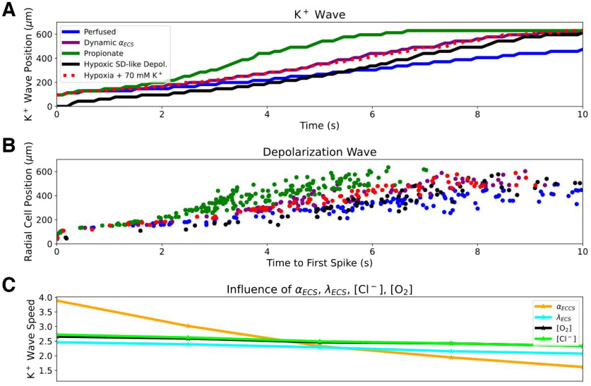
Figure 5.
Hypoxia, propionate, and dynamic ECS increased SD speed principally through αECS reduction. A, Radial K+ wave position over time during SD in perfused (Movie 1), hypoxic, HSD (Movie 2), propionate conditions, and with dynamic changes in . Hypoxia, propionate, and dynamic changes in αECS facilitated propagation. B, Radial position of SD wave represented by time to first spike in 126 selected cells at different distances from center. C, K+ wave speeds with individual parameter changes (Fig. 2). αECS had the greatest impact on SD speed over a physiologically plausible range (x-axis ranges: [O2] = 0.01–0.1 mm; αECS = 0.07–0.42; λECS = 1.4–2.0; [Cl–]ECS:[Cl–]I = 3.0:65.0–6.0:130.0 mm).

