
Figure 7.
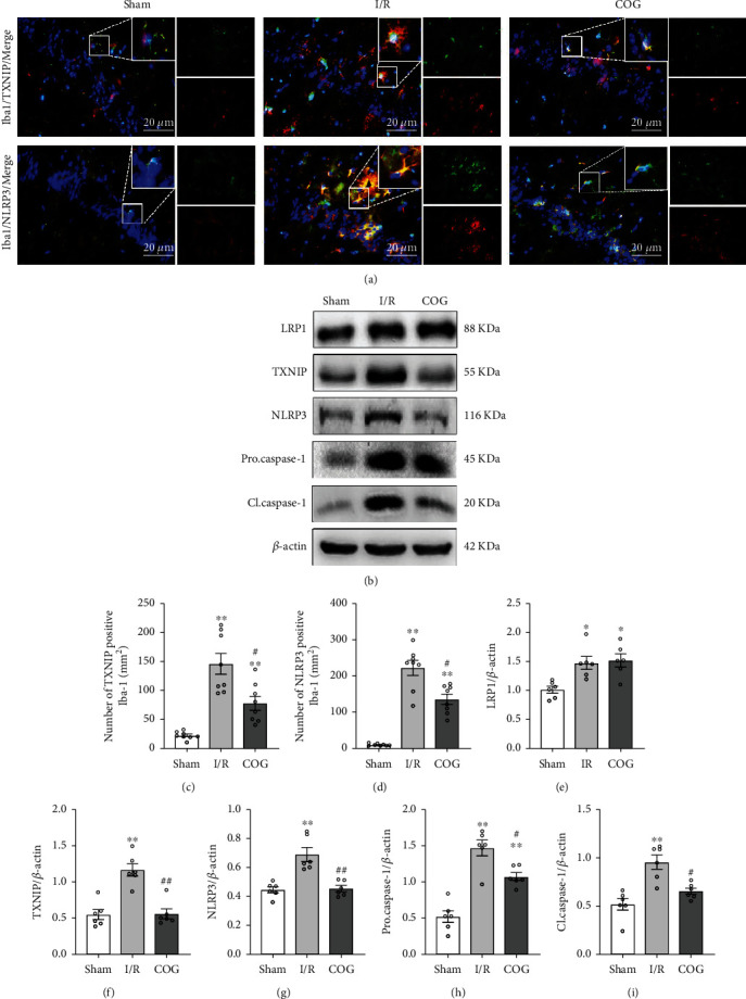
Effects of COG1410 on the TXNIP/NLRP3 signaling pathway at 72 h after cerebral I/R. (a) Representative images of the colocalization of TXNIP and NLRP3 (red) with microglia (Iba-1, green) in the hippocampal area. (b) Representative bands of Western blot data. (c, d) Quantitative analysis of TXNIP-positive and NLRP3-positive microglia. (e–i) Quantitative analysis of the Western blot bands. Data were represented as mean ± SEM. Western blot, n = 6 per group; double immunofluorescence staining, n = 8 per group. ∗P < 0.05, ∗∗P < 0.01 vs. sham group; #P < 0.05, ##P < 0.01 vs. I/R + Vehicle group.