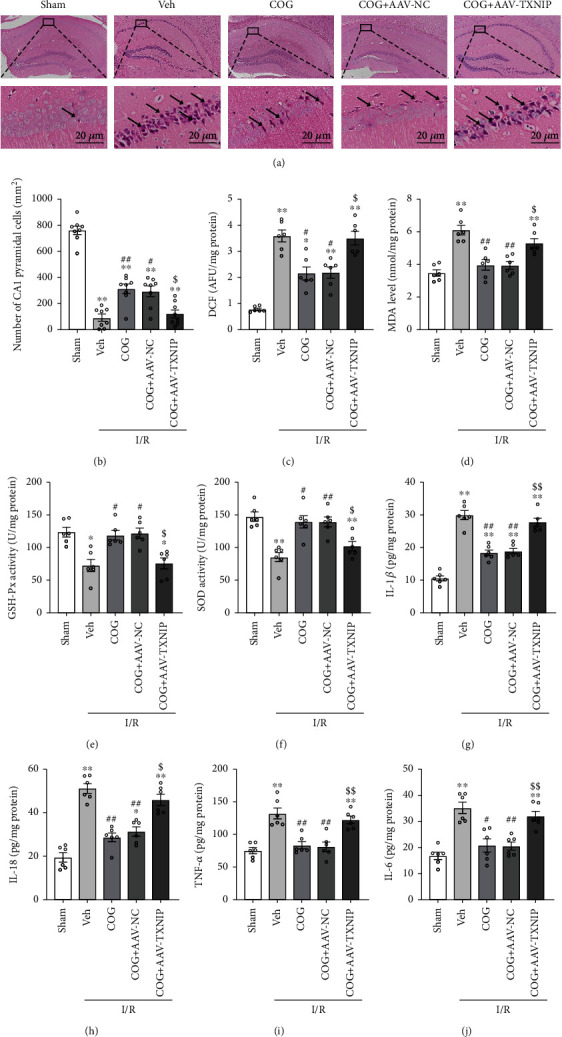
Figure 9.

Overexpression of TXNIP reversed the beneficial effects of COG1410 on histopathological damage, oxidative stress, and inflammation at 72 h after cerebral I/R. (a) HE staining of hippocampal CA1 area (×400), scale bar = 20 μm; the arrows indicate injured neurons. (b) Quantitative analysis of normal neurons in the hippocampal CA1 area. (c–f) Quantitative analysis of the oxidative stress markers (ROS, MDA, GSH-Px, and SOD). (g–j) Quantitative analysis of the inflammatory factors including IL-1β, IL-18, TNF-α, and IL-6. Data were represented as mean ± SEM. HE staining, n = 8 per group; oxidative stress markers and inflammatory factors, n = 6 per group. ∗P < 0.05, ∗∗P < 0.01 vs. sham group; #P < 0.05, ##P < 0.01 vs. I/R + Vehicle group; $P < 0.05, $$P < 0.01 vs. I/R + COG1410 + AAV-NC group.
