
Figure 2.
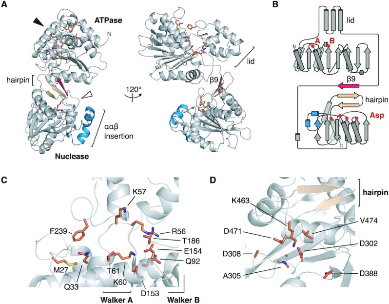
Crystal structure of the HK97 TerL. (A) Ribbon representation. Secondary structure elements unique to the HK97 protein are colored in red and blue. The conserved β-hairpin of viral terminase nucleases is colored in beige. The putative ATPase and nuclease active sites, depicted in (C) and (D), are indicated with a black and white arrowhead, respectively, with important residues shown as sticks. (B) Topology diagram of the HK97 protein. The location of the Walker A, B residues, and aspartate residues in the nuclease active site are marked. Close-up view of the (C) ATPase and (D) nuclease active sites.