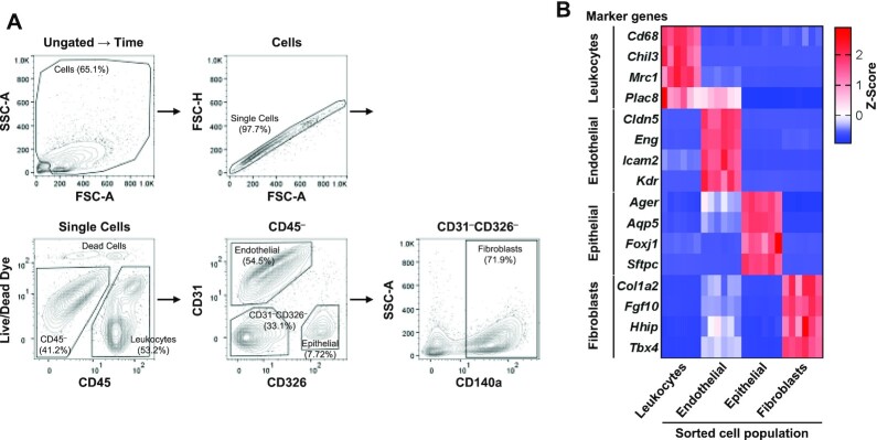
Figure 2.
Flow cytometry analysis strategy to identify leukocytes, endothelial cells, epithelial cells, and fibroblasts of mouse lung. Enzymatically dissociated mouse lungs were stained with VioGreen-CD45, APC-CD31, FITC-CD326, and PE-Vio770-CD140a antibodies, and SYTOX Blue Live/Dead cell dye. (A) After time gating, debris and non-single cells were excluded by gating on FSC-A × SSC-A and FSC-A × FSC-H. The cell population of different cell types was separated as follows: leukocytes (CD45+), endothelial cells (CD45–CD31+CD326–), epithelial cells (CD45–CD31–CD326+), and fibroblasts (CD45–CD31–CD326–CD140a+). A ratio of each cell population to the parent population was presented. (B) Each cell population was collected using the fluorescence-activated cell sorter, and RNA was isolated. RNA sequencing analysis revealed the enriched gene expression of cell-type-specific markers in each cell type. FSC, forward scatter; SSC, side scatter; A, area; H, height.

