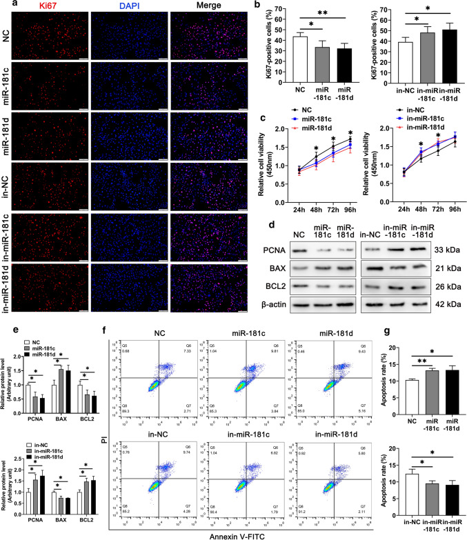
Fig. 3.
miR-181c/d inhibits proliferation and promotes apoptosis of murine Sertoli cells. The murine SCs were transfected with mimics NC, miR-181c/d mimics, inhibitors NC, or miR-181c/d inhibitors. miR-181c inhibitors, miR-181d inhibitors, and inhibitors NC are abbreviated to in-miR-181c, in-miR-181d, and in-NC, respectively. a Immunofluorescence staining of the cell proliferation marker Ki67 (red) in miR-181c/d mimics or inhibitors treated murine SCs. Scale bar: 100 µm. b Quantification of Ki67-positive cells in miR-181c/d mimics or inhibitors treated murine SCs. c CCK-8 assay performed in miR-181c/d mimics or inhibitors treated murine SCs. d Western blot analysis of PCNA, BAX, and BCL2 in miR-181c/d mimics or inhibitors treated murine SCs. The quantification of protein level is shown in the bar graph (e). f Annexin V-FITC/PI and flow cytometry analysis was used to examine cell apoptotic rate in miR-181c/d mimics or inhibitors treated murine SCs. g Quantification of cell apoptotic rate in miR-181c/d mimics or inhibitors treated murine SCs. Data are presented as mean ± SD of at least three independent experiments. *p < 0.05; **p < 0.01

