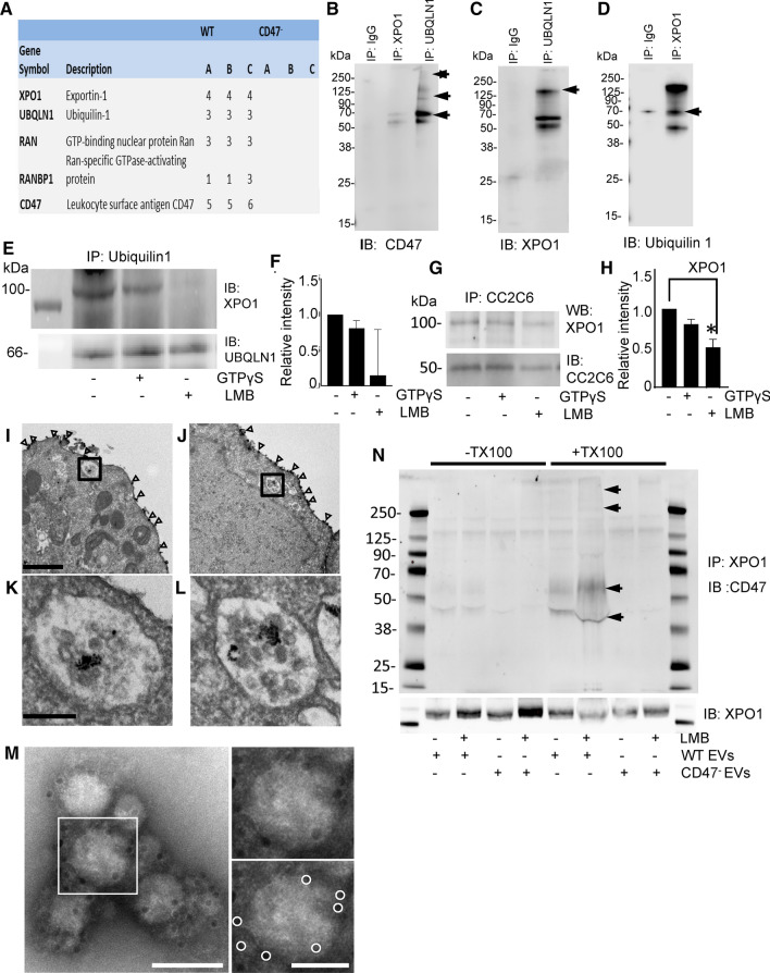
Fig. 2.
CD47 localization in EVs and interactions with the Ran/exportin-1 complex. a Cell lysates from WT and CD47− Jurkat cells were immunoprecipitated with streptavidin beads using a biotin-conjugated CD47 antibody (CC2C6). Proteins eluted from the beads were digested and subjected to LC–MS analysis to identify CD47-interacting proteins. A is the number of unique peptides, those peptides with unique sequence that are only found in the given protein. B is the number of total peptides, which is those with unique sequence that are found in the given protein but may also be found in other proteins. C is the number of peptide spectral matches, which is the total number of MS/MS spectra representing peptides from the given protein, including redundant spectra for the same peptide sequence. b–d The interactions identified by LC–MS were validated using immunoprecipitation of CD47, ubiqulin-1 and exportin-1. e Representative blot showing effects of LMB and GTPγS treatment on immunoprecipitation of exportin-1 with ubiquilin-1 in Jurkat whole cell lysates f Quantitative analysis of 3 replicate experiments with exportin-1 protein density normalized to ubiquilin-1. g, h Representative exportin-1 immunoblot and quantification of three replicate experiments (H) showing effects of LMB and GTPγS treatment on the association of exportin-1 with CD47 immunoprecipitated using CC2C6, p = .033 (*), i–l Electron micrographs for immunogold labeling to localize CD47 in vehicle treated i, k and LMB treated (J.L) Jurkat T cells. Open arrowheads indicate CD47 in the plasma membrane. Boxed areas in I and J, enlarged in K and L, respectively, show intracellular labeling for CD47 localized to MVBs. Scale bar for I-J indicates 1 μm; scale bars for K-L indicate 200 nm. m CD47 immunogold labeled and negative stained electron micrograph of EVs released by WT T cells and isolated using Exo-spin. Boxed area around one EV is shown enlarged to the right (top), with immunogold particles circled in white below. Scale bars indicate 100 nm (left), and 50 nm (right). n Immunoprecipitation of exportin-1 was performed using lysates (± Triton-X100) of EVs derived from WT and CD47− cells or cells treated with 20 nM LMB. Western blotting was performed to detect CD47 (glycosylated isoforms indicated by arrows) and exportin-1

