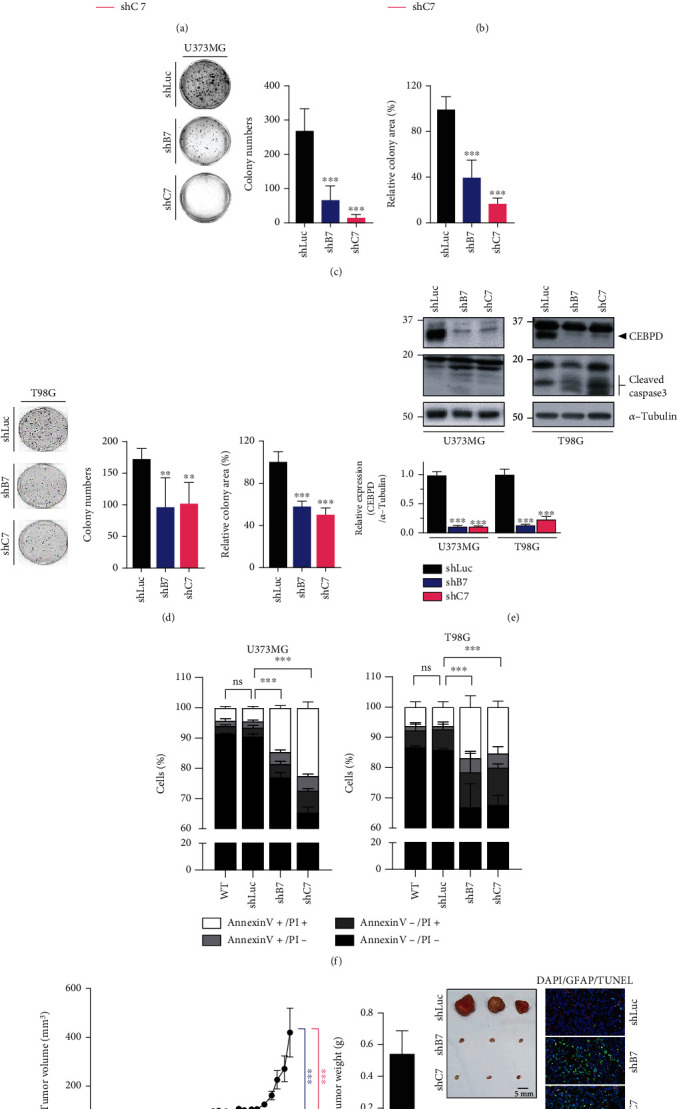
Figure 1.

Loss of CEBPD attenuates cell viability and induces cell apoptosis in GBM. (a, b) Knockdown of CEBPD reduces GBM cell viability. Cells from U373MG or T98G stable clones were subjected to CCK-8 proliferation assays. (c, d) Stable knockdown clones of (c) U373MG or (d) T98G cells were subjected to colony formation assays and grown for 7 days. The quantitative results of colony numbers and size are shown in the middle and right panels. (e) Attenuated CEBPD increases cleaved caspase 3 expression in GBM cells. Western blot analyses were conducted with the indicated antibodies using protein lysates from U373MG or T98G stable clones. Expression of α-tubulin served as the internal control. Lower panel shows the quantification of CEBPD protein expression. (f) Cells were harvested from U373MG or T98G stable clones and stained with Annexin V and Propidium Iodide (PI) for flow cytometry analysis. (g) Cells (2 × 10⁶) from stable T98G clones were injected subcutaneously into NOD-SCID mice. The mouse brain was paraffin embedded and subjected to histological analysis (right panel). Brain slides were stained by GFAP antibody and TUNEL apoptosis assay and photographed by microscope. Bars represent the means ± SEM from three independent experiments. Differences among groups were determined with one-way or two-way ANOVA followed by Tukey's multiple comparison test. ∗∗∗p < 0.001 and ∗∗p < 0.01. ns: no significant; shLuc: shRNA for luciferase; shB7, shC7: shRNAs for CEBPD.
