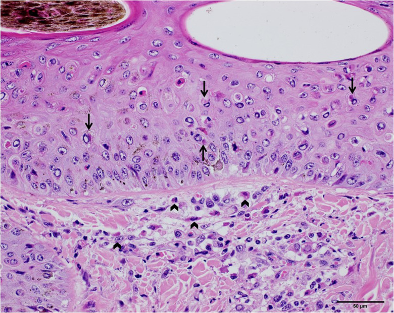
Fig. 3.

The skin of the diseased cattle (Cattle ID: 104V1290). Hydropic degeneration was observed in the dermis, as showed in the upper half of the photograph. Eosinophilic intracytoplasmic inclusion bodies in the keratinocytes and macrophages were pointed with arrows (scale bar=50 µm). The inclusion bodies in the fibroblasts and macrophages were pointed with wedge symbols.
