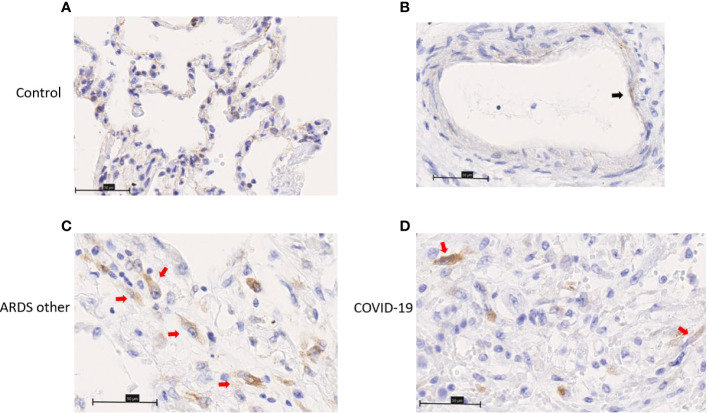
Figure 5.
Immunohistochemistry staining of chemerin on lung slides from autopsied COVID-19 patient, patient with ARDS from another origin (ARDS other) and control. (A) Representative image of alveolar lining staining. (B) Representative image of endothelial cell (black arrow) staining. (C, D) Representative image of chemerin staining of spindle cell (red arrows) in organizing phase of diffuse alveolar damage. Field magnification 400x. Scale bar: 50µm.

