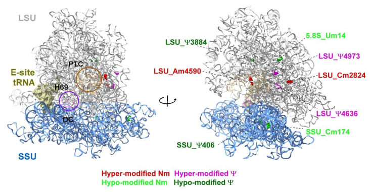
Figure 9.
Localization of Ψ and Nm residues altered in KSHV-infected iSLK cells undergoing lytic reactivation. Uninfected SLK cells and BAC16-infected iSLK cells were treated with Dox and n-Butyrate for 48 h. The data are derived from HydraPsiSseq and RiboMeth-seq analysis. The location of peptidyl transferase center (PTC), E site tRNA, decoding center (DC) and helix H69 are shown. The large subunit (LSU) and small subunit (SSU) are colored in grey and blue, respectively. The 3D representation of human ribosome is based on the published CryoEM structure (PDB 6EK0) [98]. Figures were generated using UCSF Chimera-X software (https://www.rbvi.ucsf.edu/chimerax, accessed on 16 July 2022).

