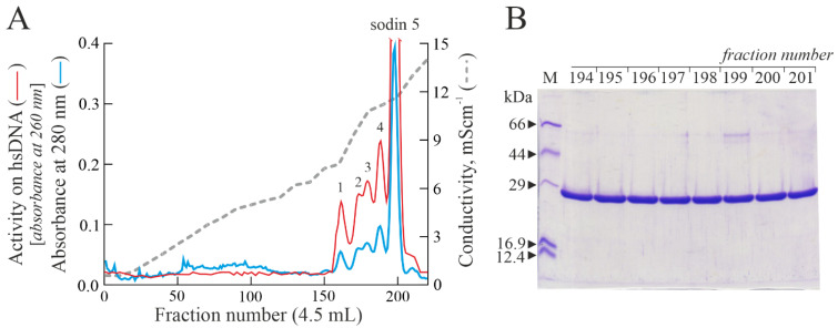
Figure 1.
(A) Elution profile after cation exchange chromatography on the CM-Sepharose column, showing five peaks (peaks 1–4 and sodin 5) with PNAG activity (arbitrary units). (B) SDS-PAGE analysis of 194–201 fractions (5.0 μg) from sodin 5 obtained after cation exchange chromatography (A). M, molecular weight markers. SDS-PAGE in the presence of β-mercaptoethanol was carried out in 12% polyacrylamide separating gel and then stained with Coomassie brilliant blue.

