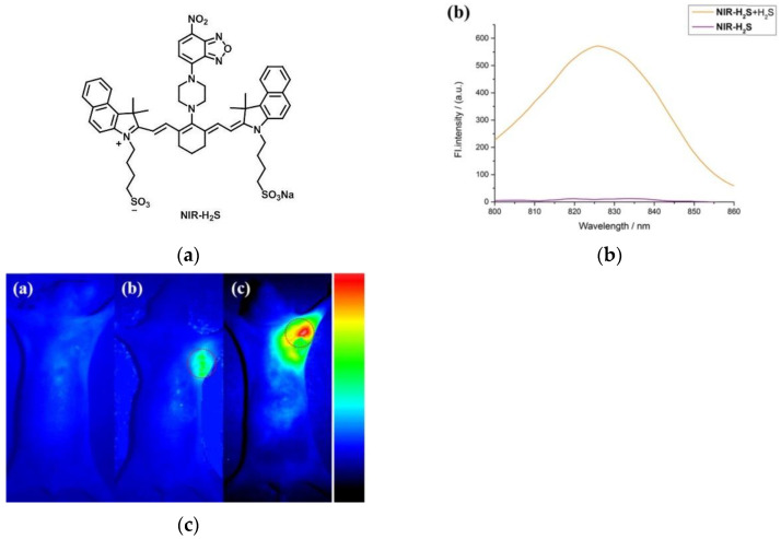
Figure 28.
(a) Structure of probe NIR-H2S; (b) Fluorescence spectra of probe NIR-H2S; (c) Fluorescence images of the tumor-bearing nude mice (c-a) The normal, (c-b) the HepG2 tumor-bearing and (c-c) the MCF-7 tumor-bearing nude mouse was injected with NIR-H2S. Reproduced with permission from [121]. Copyright 2018 Elsevier B.V.

