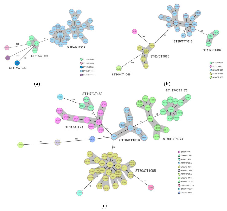
Figure 2.
Genotyping results of VRE isolates from a nosocomial outbreak in two interconnected hospitals in Southern Germany, October 2015–November 2019. (a) Core genome multi-locus sequencing typing at the beginning of the outbreak 2015–2016. The large cluster of ST80/CT1013 is highlighted in blue. (b) Genotype distribution of isolates obtained from a point prevalence study in May 2017. (c) Genotypic assessment of isolates obtained between December 2018 and March 2019. Color-coding according to complex types (CT) based on the cgMLST nomenclature. Sequence types (ST) and CTs are shown adjacent to the clusters, highlighted in grey, which represent closely related isolates (difference ≤ 15 alleles). Numbers adjacent to connecting lines represent allelic differences between isolates with variation in more than 15 alleles.

