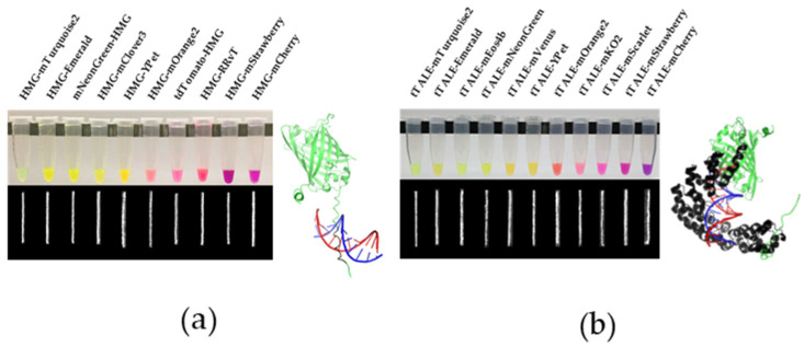
Figure 1.
Expressed FP-DBPs in test tubes and their stained DNA images. (a) HMG-FP/FP-HMG (b) tTALE-FP. FP-DBP stained λ phage DNA images were given below. Illustrations of FP-DBP binding to DNA were shown for comparison of the size of DBPs. HMG-Emerald and tTALE-Emerald structures were modeled through AlphaFold 2.1.0 (DeepMind, London, UK) [36]. DNA-binding protein motifs were colored black and were aligned to DNA by using PDB 2EZD (a) PDB 4OTO (b) in the software PyMOL.

