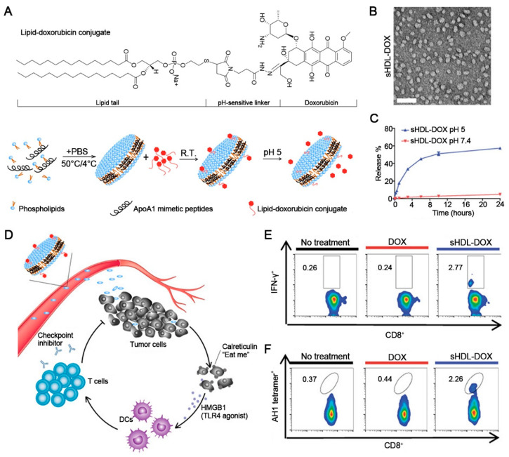
Figure 4.
Schematic of sHDL-Dox for chemo-immunotherapy. (A) Schematic of the lipid-DOX conjugate and the preparation process. (B) The image of sHDL-DOX using a transmission electron microscope. Scale bars, 50 nm. (C) The release of DOX at pH 5 and pH 7.5. Data represent mean ± SD (n = 3). (D) Schematic of sHDL-DOX triggering antigen release for cancer immunotherapy. (E) The increased percentage of IFN-γ + CD8+ T cells induced by sHDL-DOX. (F) The increased percentage of CT26 tumor antigen peptide AH1-specific CD8+ T cells induced by sHDL-DOX. Reprinted with permission from Ref. [123]. Copyright 2018, copyright the American Association for the Advancement of Science.

