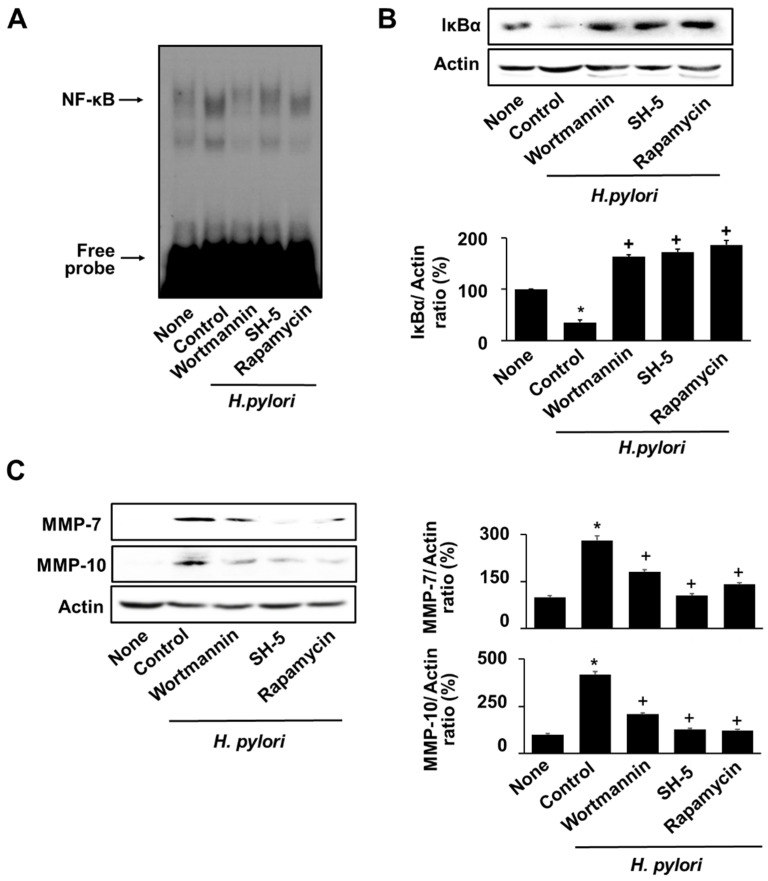
Figure 6.
Inhibitors of PI3K, AKT, and mTOR suppressed NF-κB activation, IκBα degradation, and MMPs expression in H. pylori-infected cells. 0.1 µM wortmannin, 1 µM SH-5, or 0.1 µM rapamycin was pretreated to the cells. After 2 h, H. pylori was treated to the cells and cultured for 8 h. (A) DNA-binding activity of NF-κB was measured via EMSA. (B) Protein levels of IκBα were measured via western blot analysis. Actin was used as a loading control (upper panel). The densitometry data represent the mean ± standard error (SE) from three immunoblots and are shown as the relative density of protein bands normalized to actin (lower panel). (C) The protein levels of MMP-7 and MMP-10 were determined using western blot analysis. Actin was used as a loading control (left panel). The densitometry data represent the mean ± standard error (SE) from three immunoblots and are shown as the relative density of protein bands normalized to actin (right panel). * p < 0.05 vs. ‘None’ (uninfected cells without inhibitor treatment); + p < 0.05 vs. ‘Control’ (infected cells without inhibitor treatment).

