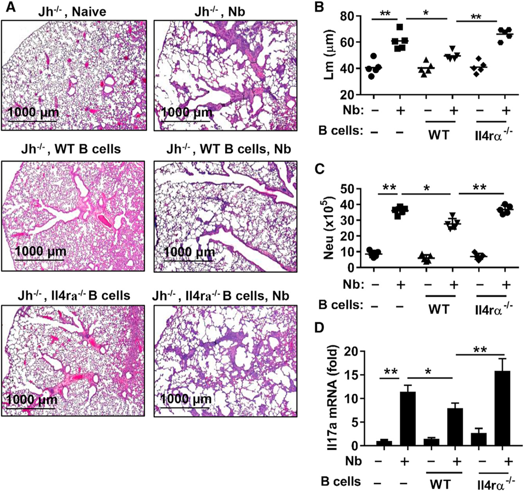
Figure 5. IL-4R Signaling Promotes B Cell-Mediated Control of Emphysema.

B cells from WT and Il4ra−/− mice were transferred to Jh−/− recipients at days −3, 0, and +1 during N. brasiliensis inoculation. Lungs were collected for analysis 7 days after inoculation.
(A) H&E staining of formalin-fixed lung sections.
(B) Emphysematous pathology was quantitated by digital imaging analysis of mean linear intercept measurements (Lm) of alveolar spaces.
(C) Number of lung neutrophils (CD11b+Ly6G+) was determined by flow cytometry. Each symbol represents an individual mouse, and horizontal lines indicate the mean.
(D) Lung tissues were examined for the expression of Il17a by qPCR. Gene expression is presented as the fold increase over naive WT mice after normalization to 18sRNA and expressed as the mean and SEM from five individual mice per group. Data shown are representative of at least two independent experiments (*p < 0.05, **p < 0.01). See also Figure S3.
