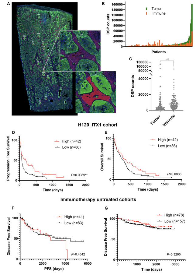
Figure 2.
Validation of CD44 expression in the tumor compartment as an indicative biomarker of sensitivity to single-agent PD-1 axis blockade in NSCLC. (A) Representative image of a whole tissue section with 12 ROIs selected from immune-enriched (panCK+/CD45+) intratumoral areas using DSP. Fluorescence image is shown on the top, and the compartmentalized image at the bottom; panCK (green), CD45 (red), SYTO13 (blue). (B), Dynamic range of CD44 expression in the tumor compartment (panCK+) and in the immune compartment (panCK–/CD45+) using DSP; (C) Comparative analysis of CD44 levels measured by DSP in the tumor compartment and in the immune compartment. (D–E) Kaplan-Meier PFS curve (D) and OS curve (E) according to CD44 expression in the tumor compartment using DSP (tertile cutpoint). (F–G) Kaplan-Meier disease-free survival curves according to CD44 expression using DSP (F) or QIF (G) in immunotherapy untreated cohorts (CIMA-CUN cohort and YTMA423 cohort as F and G, respectively) (tertile cutpoint). CK, cytokeratin; DSP, digital spatial profiling; ns, not significant; NSCLC, non-small-cell lung cancer; OS, overall survival; PD-1, programmed cell death protein-1; PFS, progression-free survival; QIF, quantitative immunofluorescence; ROI, regions of interest.

