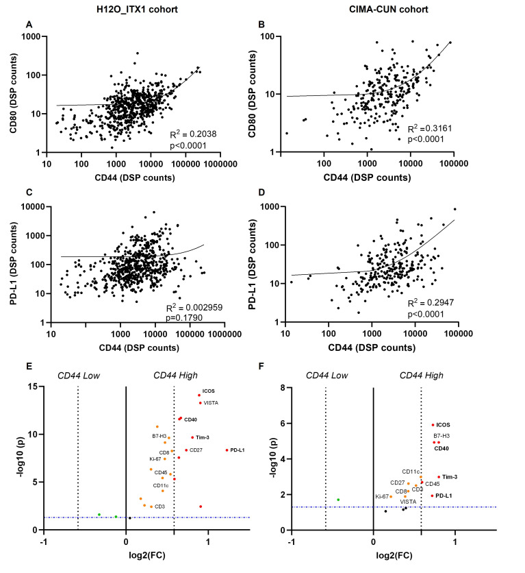
Figure 3.
CD44 levels in the tumor compartment and immune microenvironment features in human NSCLC. (A–B) Correlation between CD44 and CD80 levels in the tumor compartment in H12O_ITX1 cohort (A) and CIMA-CUN cohort (B). (C–D) Correlation between CD44 and PD-L1 levels in the tumor compartment in H12O_ITX1 cohort (C) and CIMA-CUN cohort (D). (E–F) Differentially expressed protein markers in ROIs with high CD44 expression in the tumor compartment (top tertile) relative to ROIs with low CD44 expression in the tumor compartment (rest) in H12O_ITX1 cohort (E) and CIMA-CUN cohort (F). The significance (FDR-adjusted p values) is represented relative to the FC in protein levels in CD44 high relative to CD44 low ROIs. Only statistically significant markers are highlighted. Markers with a FC >1.5 and FDR-adjusted p values<0.05 in the two cohorts are marked in red bold. DSP, digital spatial profiling; FC, fold change; FDR, false discovery rate; PD-L1, programmed death-ligand 1; NSCLC, non-small-cell lung cancer; ROI, regions of interest.

