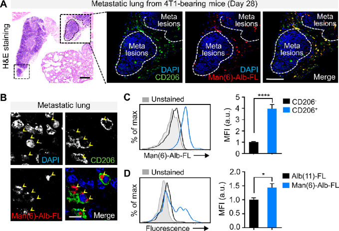
Figure 5.
Confirmation of Man(6)-Alb-FL as a macrophage-targeted probe. (A) Representative H&E stained images of lungs from 4T1-bearing mice injected with Man(6)-Alb-FL 28 days after tumor injection (left). Representative confocal immunofluorescent images showing in vivo colocalization of injected Man(6)-Alb-FL (red) with the CD206+ macrophages (green) within the lungs from 4T1-bearing mice, as confirmed in the overlay image (yellow). The white dotted lines indicate metastatic nodules in the lung. Scale bar = 75 μm. Higher magnification view (B) further demonstrates CD206+ macrophage-specific uptake (yellow arrowheads) in vivo. DAPI, blue. Scale bar = 10 μm. (C,D) Quantification of CD206+ macrophage-specific uptake in vivo using flow cytometry. (C) Man(6)-Alb-FL uptake by each gated CD206– macrophage (CD45+CD11b+F4/80+CD206–) and CD206+ macrophage (CD45+CD11b+F4/80+CD206+) subset was determined from the fold change in the mean fluorescence intensity (MFI) of colabeled fluorescent dye (FNR-648). (D) Fold change in MFI of colabeled FNR-648 on CD206+ macrophages from 4T1-bearing mice following injection with Alb(11)-FL or Man(6)-Alb-FL. n = 4–7 mice/group. Data represent the mean ± SEM. *P < 0.05, ****P < 0.0001 using Student’s t test.

