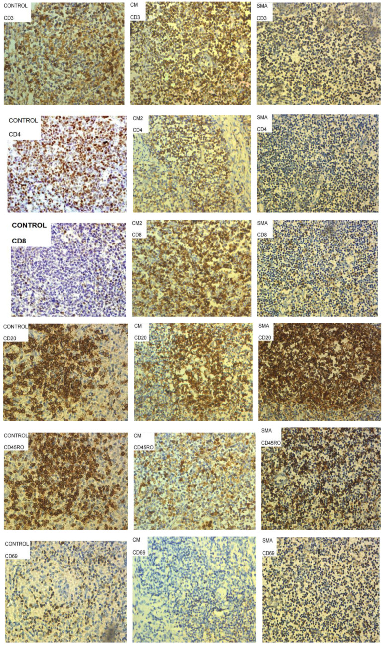
Figure 2.
Representative images of lymph node tissue sections from patients who died of sepsis (controls), Severe Malarial Anemia (SMA) and Cerebral Malaria (CM) stained with antibodies to characterize CD4+ T cells, CD8+ T cells, CD20+ B cells, activated cells (CD69+) and memory cells (CD45RO+). Cells stained brown were considered positive for the marker of interest, and representative examples are circled and highlighted by arrows in some panels above.

