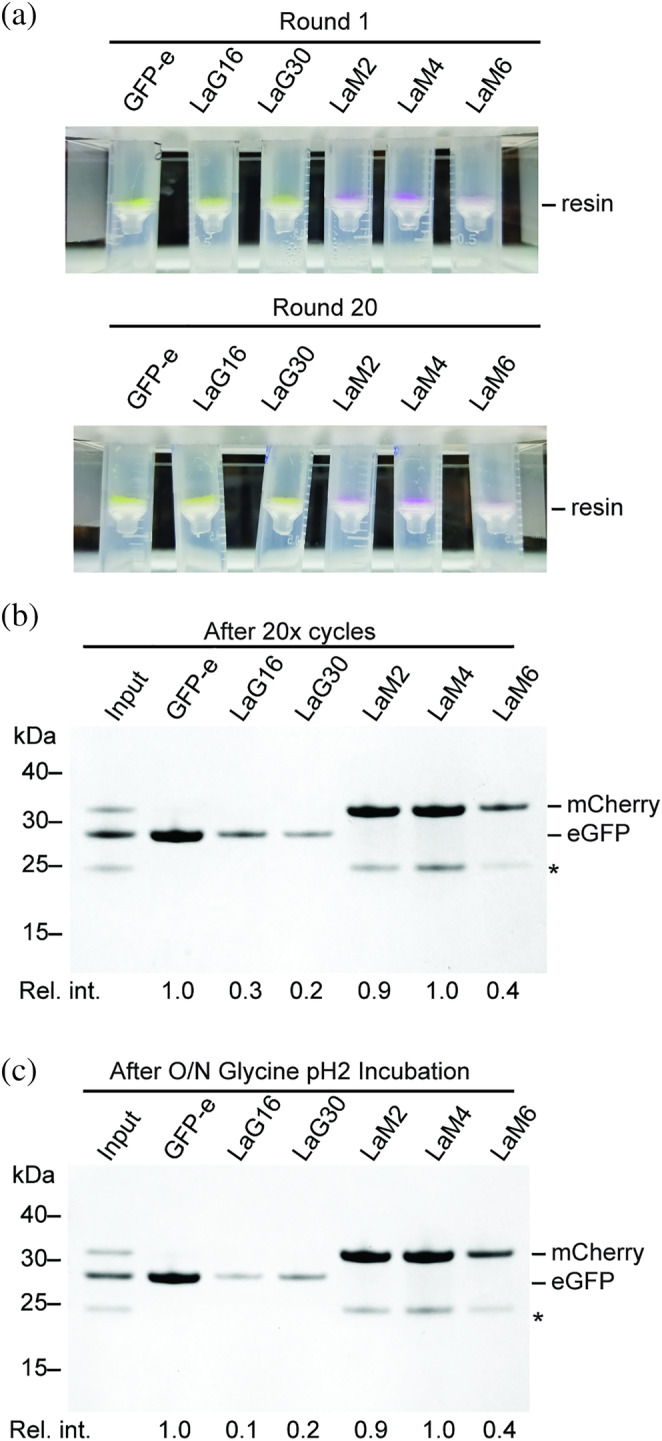
FIGURE 5.

Stability of nanobody sepharose resins to regeneration at low pH. (a) Nanobody‐sepharose resins after binding to saturating amounts of FP in the initial round, or after 20 cycles of binding and regeneration. Binding of FP to resin can be observed visually on the spin filter where the resin accumulates. GFP‐e is GFP‐enhancer nanobody. (b) SDS‐PAGE of protein bound to resin in Panel (a) after 20 cycles of binding and regeneration. This is comparable to the first cycle of binding as shown in Figure 3a. *Band corresponding to degraded mCherry. (c) SDS‐PAGE analysis of protein bound to nanobody‐sepharose resins that had been pre‐incubated in glycine pH 2 for overnight. Relative intensity of bound FP is indicated below each lane
