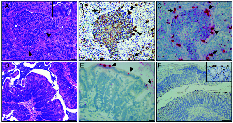
Figure 2.
Histopathology of the lung and large intestine from a 1-y-old, female, NSG mouse. (A) Representative airway demonstrating a bronchiolar and alveolar inflammation characterized by luminal neutrophilic infiltration mixed with necrotic debris and proteinaceous material. Multifocally, bronchiolar epithelial cells exhibit intracytoplasmic clear vacuoles with pale-basophilic structures compatible with Chlamydial inclusions (arrowhead and inset). Peribronchiolar and alveolar space is infiltrated with moderate numbers of macrophages and neutrophils intermixed with reactive fibroblasts (scale bar = 50 µm). (B) IHC of the lung demonstrating detection of chlamydial MOMP antigen in bronchiolar epithelial cells (arrowhead; brown staining) and areas with peribronchiolar inflammation (arrow, scale bar = 50 µm). (C) ISH demonstrates positive staining (red) for Cm mRNA in bronchiolar epithelial cells (arrowhead) and areas of peribronchiolar inflammation (arrow; scale bar = 50 µm). (D) Representative H and E-stained section of a normal cecal wall (scale bars = 100 µm). (E) High magnification field demonstrates ISH signal (red staining) in the cecal epithelium (arrow) and lumen (arrowhead; scale bar = 50 µm). (F) IHC of descending colon demonstrating detection of intracytoplasmic chlamydial MOMP antigen in surface epithelial cells (inset - brown staining; scale bars = 20 and 200 µm).

