Figure 4.
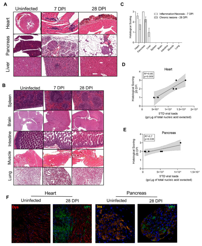
Histological lesions and cell dysfunctions in organs of CVB3/28-infected DBA/2J mice. (A) In the infected group, cardiac lesions were found from 7 DPI to 28 DPI. Infected mice exhibited multiple inflammatory foci at 7 DPI. At 14 and 28 DPI, calcified fibrosis was found. At 7 DPI, diffuse pancreatic inflammatory infiltrates with necrosis were found, sparing islets cells. After 14 DPI, an adipose tissue replacement was found, sparing islets cells. Hepatitis was found at 7 DPI, characterized by sparse inflammatory and necrosis foci or sub capsular inflammatory infiltrates. After 14 DPI, no inflammatory or scarring lesions were found. HES, haematoxylin-eosin-safran staining. Original magnification: 10× to 400×. (B) Neither acute nor chronic lesions were found in other organs including the brain, spleen, intestines, lungs, or muscle. Original magnification: 10× to 400×. (C) Inflammation and necrosis were found in the heart and pancreas at 7 DPI (n = 3 to 5). Fibrosis was present only in the heart at 28 DPI (n = 4). Inflammation and necrosis were present in the liver at 7 DPI (n = 3) only, with no chronic histological lesion. No lesion was found in other organs (n = 3). Data represent mean +/− SEM. (D,E) In the heart (n = 6) and pancreas (n = 6) of infected DBA/2J mice, a positive correlation between histological lesion scoring at 28 DPI and the 5′TD EV-B load. Linear regression curve, with 95% confidence intervals in grey. (F) Confocal analyses revealed a colocalization of VP1 and dystrophin disruption in the heart, and of VP1 and a decrease in insulin staining in the pancreas, at 28 DPI. DPI: Days Post Infection.

