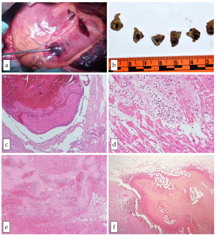
Figure 1.
Gross and microscopic cardiac features in case #1. Panel (a) shows the transmural myocardial rupture. Panel (b) shows the transversal section of the coronary artery with evidence of atherosclerotic changes and an intracoronary clot. In panels (c–f), the histological findings of the myocardial sections are presented (hematoxylin–eosin staining): a fresh thrombus in the coronary artery (c, original magnification 10×); inflammatory infiltrates (neutrophils) and myocyte contraction band necrosis (d, original magnification 20×); myocardium adjacent to the rupture site with intense inflammatory infiltrates, fibrin, and coagulative necrosis (e, original magnification 10×); hemorrhage in the epicardial adipose tissue close to the left ventricular (LV) free wall rupture (f, original magnification 20×).

