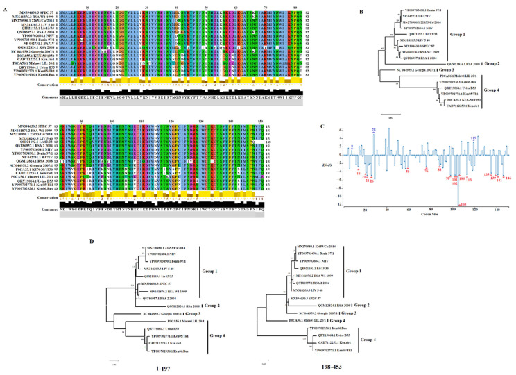
Figure 1.
Evaluation of A151R protein across ASFV isolates. (A) Amino acid alignment representing the diversity of A151R protein of ASFV in the field. Residues in white spots represent changes between amino acids with different charges. Conservation plot scores reflect the nature of the change in specific sites, with high scores associated with changes in similar biological properties. Alignment was produced using the software Jalview version 2.11.1.4. (B) Phylogenetic analysis representing the diversity of A151R protein of ASFV in the field. Based on the cluster distribution, isolates were categorized into five groups. Numbers above internal branches represent bootstrap values (1000 repetitions). (C) The graphic represents the dN (rate of evolution at non-synonymous sites), dS (rate of evolution at synonymous sites), and ratio (dN-dS) at specific codon sites in the A151R gene of ASFV. Blue and red asterisks represent codon sites evolving under diversifying and purifying selection, respectively. Analyses were conducted using the evolutionary algorithms FEL and MEME using cutoff values of p = 0.1. (D) Phylogenetic analysis showing the topology incongruence produced by different segments where the single breakpoint at nucleotide 197 was detected by GARD. Phylogenetic analysis was conducted with the maximum likelihood method, using the general time reversible model.

