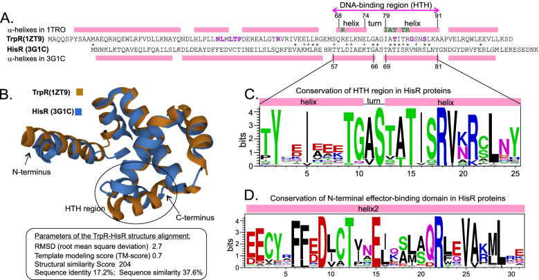
Fig. 4.
Structural analysis of HisR and TrpR regulators. A Alignment of TrpR repressor from E. coli and HisR protein from Eubacterium eligens. An asterisk marks identical residues. Pink boxes mark the position of alpha-helices in both structures. Helix-turn-helix (HTH) motif of DNA binding region is marked. Residues involved in contacts between TrpR and DNA operator within the HTH motif are shown in green (according to [28]). Tryptophan binding residues in TrpR are shown in purple (according to the 1ZT9 structure). B Pairwise structure alignment of E. coli TrpR (1ZT9) and E.eligens HisR (3G1C) obtained by the RCSB PDB web tool. C Sequence logo of the HTH motif in HisR proteins. Logo was built based on alignment of all analyzed HisR proteins from 492 genomes of Firmicutes and other lineages. Positively and negatively charged amino acids are shown in blue and red, respectively

