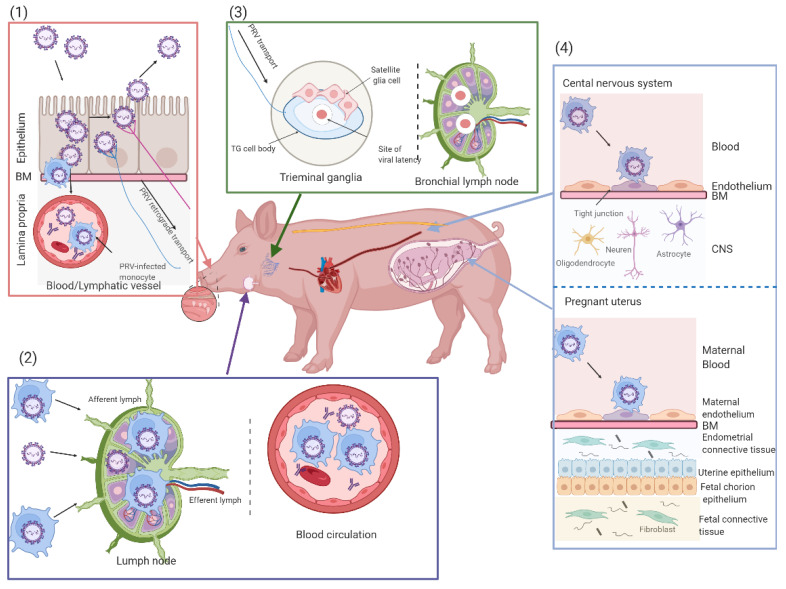
Figure 4.
Schematic representation of the pathogenesis of PRV in pigs in different stages of growth. (1) Primary viral replication in the epithelial cells (ECs) of the upper respiratory tract: PRV first infects epithelial cells, with a viral spread and shedding, and then crosses the basement membrane (BM) and lamina propria by using single infected leukocytes to reach the blood circulation and draining lymph nodes. Lastly, PRV entry occurs at nerve endings of the peripheral nervous system and diffuses retrogradely to trigeminal ganglia (TG). (2) PRV replication in the draining lymph nodes and cell-associated viremia. (3) Establishment of PRV latency in the trigeminal ganglia (TG) neurons. (4) Secondary replication in target organs (the pregnant uterus and the central nervous system (CNS)): the secondary replication in the ECs of the pregnant uterus can lead to vasculitis and multifocal thrombosis, with an abortion of sows, and in newborn piglets, sudden death usually occurs in the absence of clinical signs.

