Figure 1.
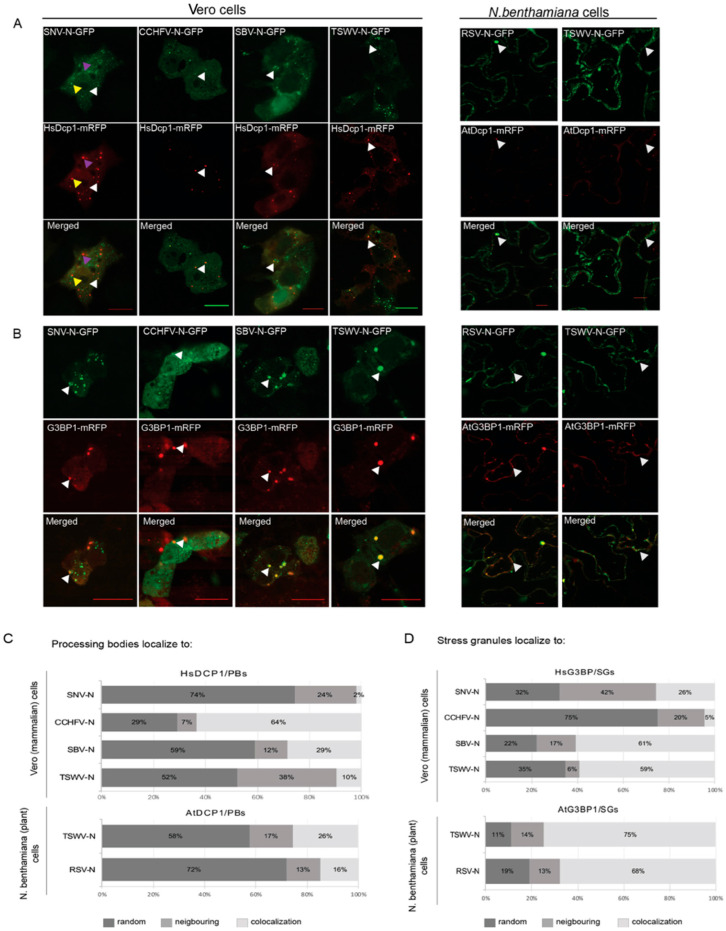
Localization and quantification of different NSV N proteins relative to PB/SG in plant cells and animal cells. (A) Fluorescence microscopy images of SNV, CCHFV, SBV and TSWV N-GFP protein localization relative to P body marker protein HsDcp1-mRFP (left panel) in Vero cells, and of RSV and TSWV N-GFP protein localization relative to P body marker protein AtDcp1-mRFP in plant cells (right panel). (B) Fluorescence microscopy images of SNV, CCHFV, SBV and TSWV N-GFP protein localization relative to SG marker protein HsG3BP1-mRFP (left panel) in Vero cells (subjected to arsenite treatment for SG-induction), and of RSV and TSWV N-GFP protein localization relative to SG marker protein AtG3BP1-mRFP in plant cells (subjected to a heat shock for SG-induction). (C) Quantification of P body localization relative to different N proteins. Colocalization, neighboring and random localization ratio were quantified for SNV, CCHFV, SBV and TSWV N protein in animal cells, and TSWV and RSV N protein in plant cells. (D) Quantification of SG localization relative to different N proteins. Colocalization, neighboring and random localization ratio were quantified for SNV, CCHFV, SBV and TSWV N protein in animal cells, and TSWV and RSV N protein in plant cells, after cells were treated for SG-induction. Bar is 10 μm.

