Fig. 5.
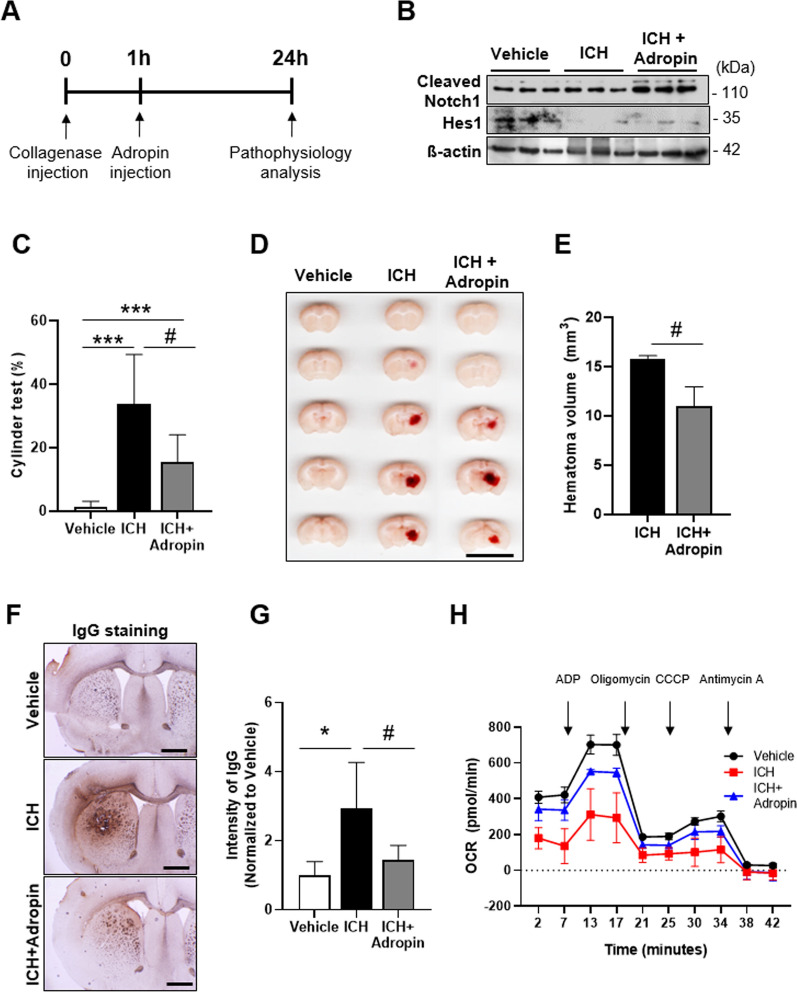
Adropin attenuates ICH pathology through activation of the Notch1 signaling pathway. A Experimental time-line for adropin injection in ICH model mice. B Attenuated Notch1 and Hes1 protein levels in the adropin-treated ICH group (n = 6 mice/group). C Cylinder test performed on ICH model mice after adropin injection (n = 11 mice/group). D Representative brain sections showing hematoma volume after adropin injection in an ICH model mouse. Scale bar: 10 mm. E Quantification of hematoma volume (n = 3 mice/group). F Representative coronal brain sections showing IgG staining after adropin injection. Scale bar: 100 µm. G IgG staining intensity, quantified using ImageJ (n = 5 mice, 8 slides per group). H Mitochondrial respiration in the striatum after adropin injection in ICH model mice, determined by OCR analysis (n = 4 mice/group). Data are presented as means ± SD from three independent experiments performed under the same conditions (*P < 0.05, **P < 0.01, ***P < 0.001, Vehicle vs. ICH; #P < 0.05, ICH vs. ICH + adropin)

