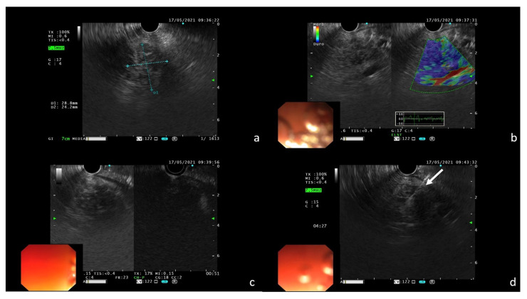
Figure 5.
EUS features of the pancreatic lesion confirming the presence of a nodular area in the pancreas body (a); dominant blue color at elastography is an indicator of high tissue stiffness (b); poor contrast enhancement after i.v. administration of SonoVue (c); FNA with 22 G needle of the nodular area ((d); white arrow).

