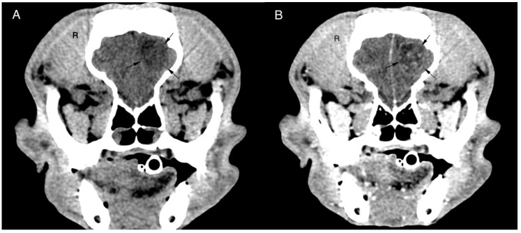
Figure 1.
Transverse pre (A) and post (B) contrast CT brain image with soft tissue algorithm. The images reveal an intra-axial, ill-defined round, hypoattenuating lesion located in the left frontal lobe of the cerebrum. The lesion shows ring enhancement after ionated contrast administration and a slight midline shift to the right. Histopathological diagnosis was oligodendroglioma grade II.

