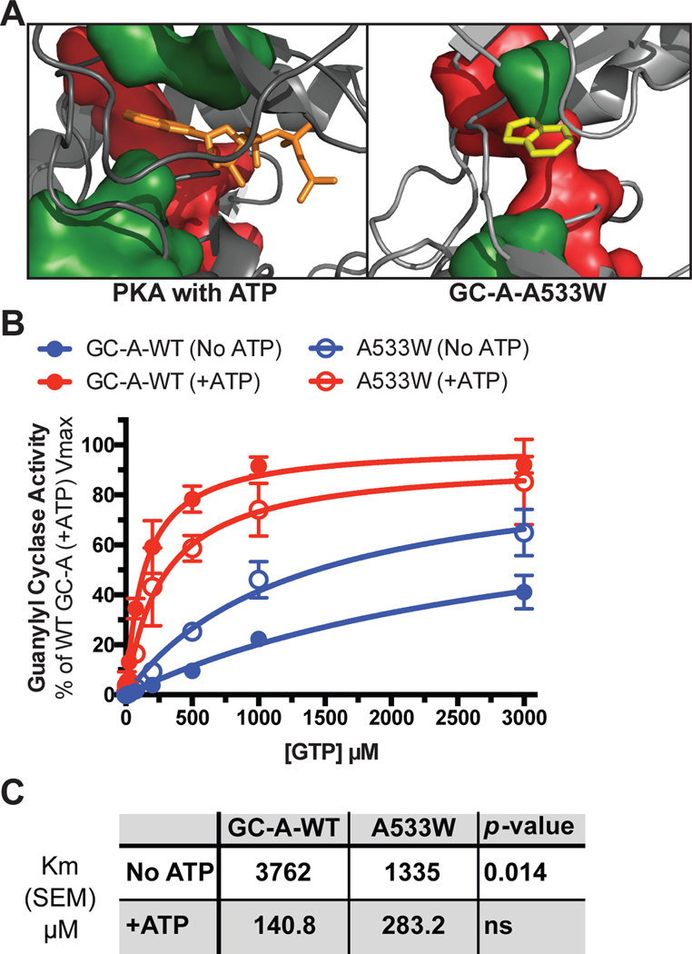
Fig. 8. The GC-A-A533W mutation partially mimics the ATP-bound state of GC-A.
(A) A model demonstrating how ATP (orange) rigidifies the C-spine (green) in PKA and how the Trp in the GC-A-A533W mutant (yellow) docks into the same pocket. The R-spine is shown in red. (B) Substrate-velocity guanylyl cyclase assays in membranes from HEK293T cells expressing WT GC-A or GC-A-A533W in the presence of MgCl2, ANP, and increasing concentrations of GTP with or without ATP. n = 4 independent experiments. (C) A table showing the measured Michaelis constants and their associated p-values. Error bars represent the SEM.

