. 2021 Jul 6;11(12):3090–3105. doi: 10.1158/2159-8290.CD-20-1741

Figure 5.

Figure 5. Activation of p53 in cancer cells enhances antigen processing and presentation. A, Heat maps show the induction of APP genes in MCF7, SJSA-1, and HCT116 cells, as assessed by RNA-seq. Absence of induction of APP genes in HCT116 p53KO cells confirms p53 dependency. B, qPCR shows the induction of APP genes in melanoma cells A375 (left) and B16 (right) upon treatment with p53-activating stapled peptide 7041 (red bars), but not control peptide 7342 (gray bars). Results shown are the mean ± SD from three independent experiments performed in triplicate (comparison between peptides vs. DMSO treatment). C, Flow cytometry shows an increased cell surface expression of HLA-A/B/C upon treatment with three MDM2 inhibitors in A375 (top) and SJSA-1 cells (bottom). Shown is propidium iodide–negative (PI−) population. D and E, p53-dependent enhanced presentation of heterologous OVA peptide-SIINFEKL on the cell surface of A375-OVA cells upon nutlin treatment, as assessed by flow cytometry (n = 3). DMSO was used as a negative control, palbociclib (Palbo) served as a positive control. F, Overexpression of FH-LSD1 prevented the induction of APP genes upon p53 activation, as assessed by qPCR in A375 cells transfected with FH-LSD1 expression vector and treated with 7041 versus control vector-transfected cells. G, Partial rescue of APP genes induction upon by ectopic expression of DNMT1, assessed as in F. B, E, F, G, Student t tests. Error bars, SD. *, P < 0.05; **, P < 0.01; ***, P < 0.001; #, P < 0.0001.

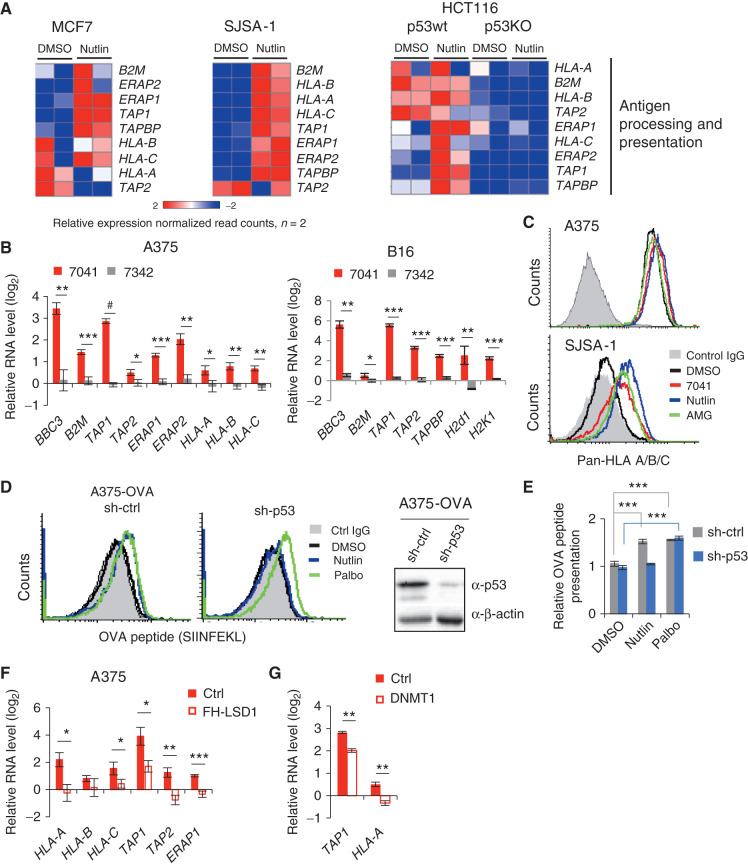
Activation of p53 in cancer cells enhances antigen processing and presentation. A, Heat maps show the induction of APP genes in MCF7, SJSA-1, and HCT116 cells, as assessed by RNA-seq. Absence of induction of APP genes in HCT116 p53KO cells confirms p53 dependency. B, qPCR shows the induction of APP genes in melanoma cells A375 (left) and B16 (right) upon treatment with p53-activating stapled peptide 7041 (red bars), but not control peptide 7342 (gray bars). Results shown are the mean ± SD from three independent experiments performed in triplicate (comparison between peptides vs. DMSO treatment). C, Flow cytometry shows an increased cell surface expression of HLA-A/B/C upon treatment with three MDM2 inhibitors in A375 (top) and SJSA-1 cells (bottom). Shown is propidium iodide–negative (PI) population. D and E, p53-dependent enhanced presentation of heterologous OVA peptide-SIINFEKL on the cell surface of A375-OVA cells upon nutlin treatment, as assessed by flow cytometry (n = 3). DMSO was used as a negative control, palbociclib (Palbo) served as a positive control. F, Overexpression of FH-LSD1 prevented the induction of APP genes upon p53 activation, as assessed by qPCR in A375 cells transfected with FH-LSD1 expression vector and treated with 7041 versus control vector-transfected cells. G, Partial rescue of APP genes induction upon by ectopic expression of DNMT1, assessed as in F. B, E, F, G, Student t tests. Error bars, SD. *, P < 0.05; **, P < 0.01; ***, P < 0.001; #, P < 0.0001.