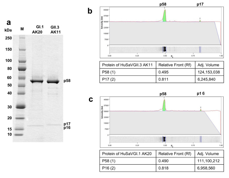
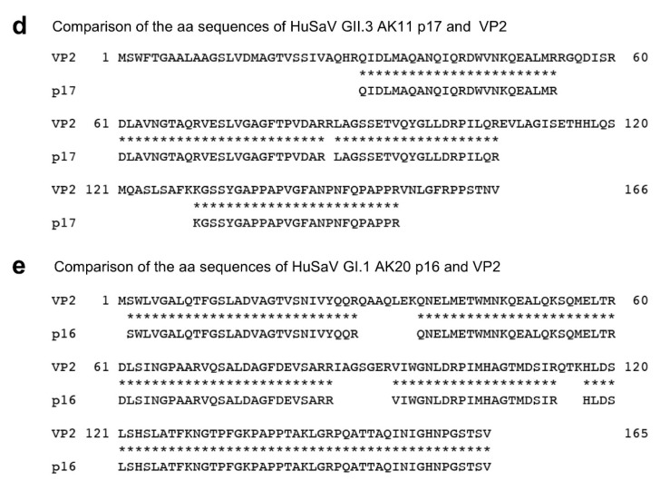
Figure 3.
Proteins analyses of the p17 and p16. Purified HuSaV GI.1 AK20 virions and HuSaV GII.3 AK11 virions were analyzed by SDS-PAGE followed by CBB staining (a). The ratio of the protein content between p58 and p17 of HuSaV GII.3 AK11 (b), and that between p58 and p16 of HuSaV GI.1 AK20 (c) were quantitated by Image software ver. 6.1 based on the band intensities. The identified aa sequences of p17 was compared with those of VP2 of HuSaV GII.3 AK11 (d), and the aa sequences of p16 was compared with those of VP2 of HuSaV GI.1AK20 (e).


