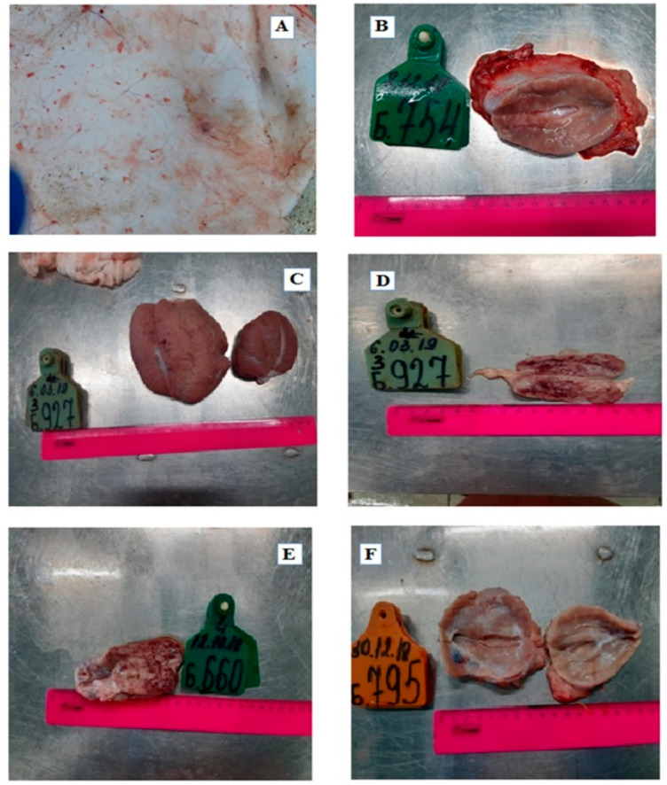
Figure 4.
Pathologies observed in various organs. (A) The hypodermis of the skin (the arrows indicate a lesion) of Bull №2; (B) Prescapular node displaying hemorrhaging, Bull №2; (C) Testes: hyperaemic, Bull №3; (D) Mesenteric lymph node, Bull №3; (E) Mesenteric lymph node, enlarged, Bull №4; (F) Posterior pharyngeal lymph node enlargement of Bull №5.

