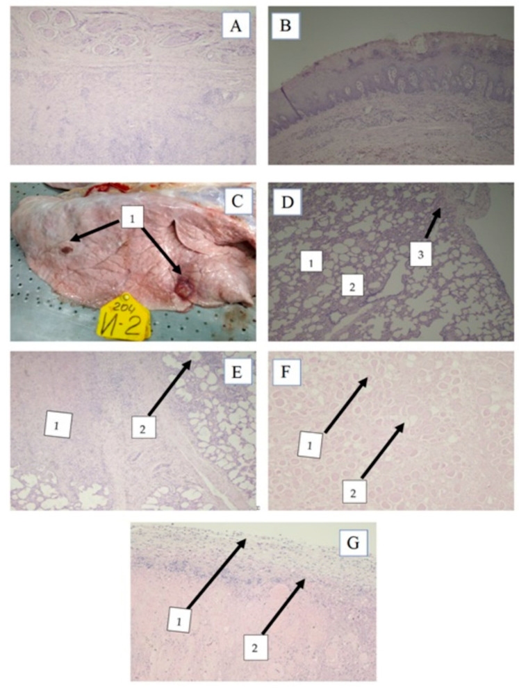
Figure 5.
Histopathologic findings of lumpy skin disease virus in cattle tissue. (A) Subacute liquefactive (pus) necrosis in the derma. H and E staining, magnification X40; (B) Esophageal Necrotic Lesion. H and E staining, magnification X40; (C) Lobular pneumonia with multiple necrotic sites: 1—foci of necrosis; (D) Affected lungs. 1—Atelectases, 2—dislectases, 3—pleurite. H and E staining, magnification X40; (E) Focal purulonecrotic pneumonia; 1—necrosis, 2—thickened interalveolar septa; (F) Muscle dystrophy. 1—edema, 2—muscle fiber dystrophy. H and E staining, magnification X40; (G) The serous membrane. Focal serositis. 1—inflammation, 2—lymphocytic infiltrations. H and E staining, magnification X4.

