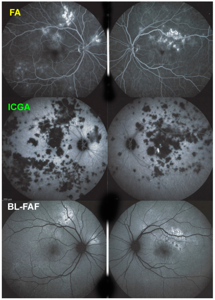
Figure 5.
APMPPE/AMIC; FA & ICGA & BL-FAF at presentation. Case of acute APMPPE/AMIC analysed by ICGA & FA & BL-FAF. This multimodal imaging shows that the initial event is choriocapillaris non-perfusion clearly shown by extensive areas of ICGA hypofluorescence (middle two frames) while retinal (FA) involvement is still limited. BL-FAF hyperautofluorescence is still limited as choriocapillaris non-perfusion induced ischaemia did not alter the outer retina yet, except in a few areas along the superior temporal arcades.

