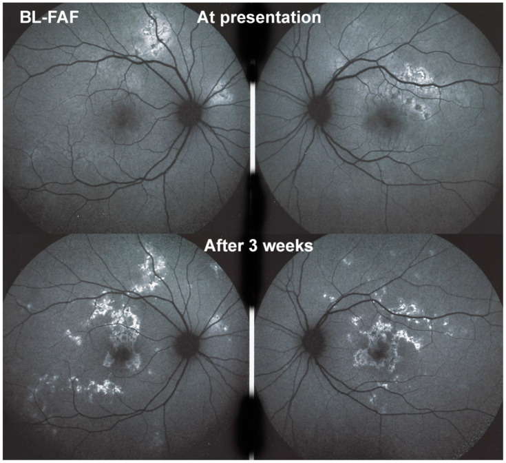
Figure 6.
APMPPE/AMIC; BL-FAF at presentation and in the subacute phase. At presentation (top two frames) there is limited hyperautofluorescence as there is mainly thickening of the outer retina due to choriocapillaris non-perfusion (see Figure 8) and limited areas (yet) of loss of photoreceptor outer segments (see Figure 7). During the subacute phase (bottom two frames) there are extended irregular areas of hyperfluorescence corresponding either to loss of photoreceptor outer segments allowing to see the hyperautofluorescent RPE lipofuscin or RPE cell damage with an accumulation of cellular fluorophore debris or both.

