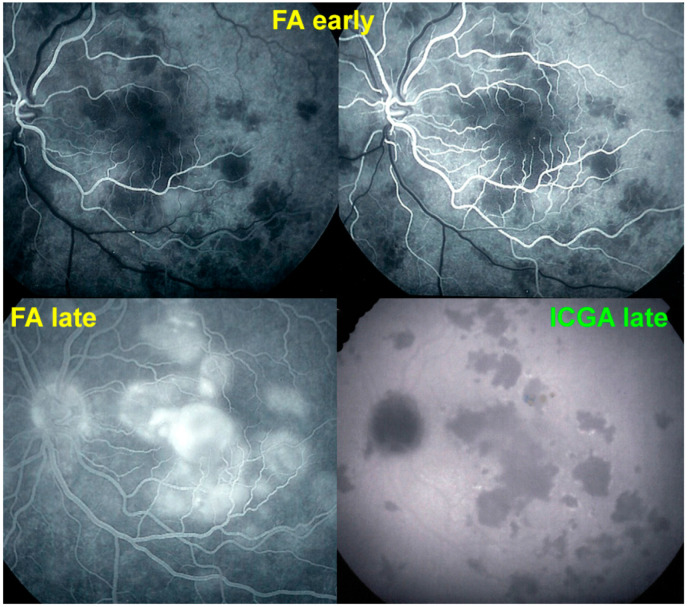
Figure 10.
Late FA pooling in a severe case of APMPPE/AMIC. In severe cases of APMPPE/AMIC, FA shows choriocapillaris non-perfusion in early angiographic frames (top two pictures—FA early) followed by areas of abundant retinal pooling in the late angiographic phase (bottom left picture—FA late). Traditionally this phenomenon is explained by the very hypothetical alleged change of polarity of the RPE and fluid movement from the choroid to the retina. However, the choriocapillaris areas under the retinal pooling are non-perfused (bottom right picture—ICGA late). Therefore, a more probable origin of the fluid is an exudation from retinal vessels in response to severe outer retinal ischemia.

